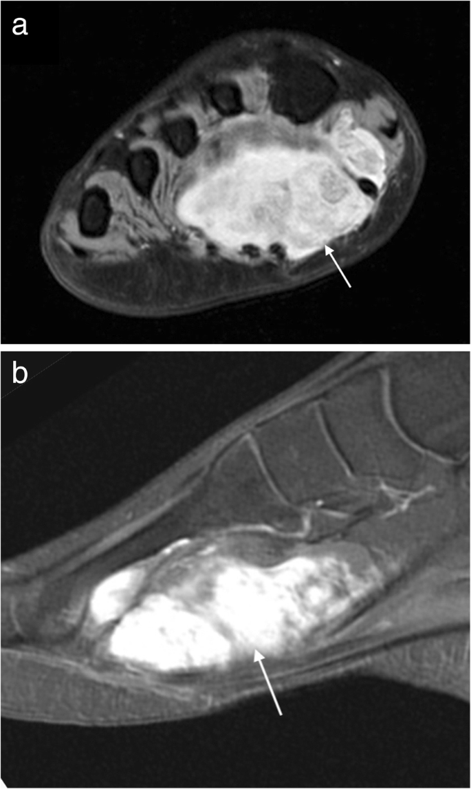
Plantar Foot Muscles Mri - Imaging Of Plantar Fascia Disorders Findings On Plain Radiography Ultrasound And Magnetic Resonance Imaging Insights Into Imaging Full Text : An mri will show a smooth, consistent (homogenous) mass that is affiliated with the plantar fascia (figure 2).
Plantar Foot Muscles Mri - Imaging Of Plantar Fascia Disorders Findings On Plain Radiography Ultrasound And Magnetic Resonance Imaging Insights Into Imaging Full Text : An mri will show a smooth, consistent (homogenous) mass that is affiliated with the plantar fascia (figure 2).. Quadratus plantae, lumbricals 3rd layer: Multiple soft tissue masses scattered in the plantar fat pad of the foot probably represent plantar fibromatosis. They are generally divided into two sets: Plantar fasciitis is an extremely common cause of heel pain. Abductor hallucis, flexor digitorium brevis, abductor digiti minimi 2nd layer:
Edited by brent brookbush dpt, pt, ms, pes, ces, cscs, acsm h/fs. Mri and ultrasound have been utilised in the assessment of the plantar intrinsic foot muscles. How does ankle mri work? Name the muscles of the plantar (sole) of the foot. Muscles of the foot are located on its rear and on the sole.
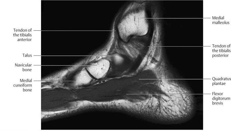
Ankle And Foot Radiology Key from radiologykey.com An mri will confirm the diagnosis and allow differentiation of other causes of masses in the foot, such. 31 the plantar intrinsic foot muscles consist of four layers of muscles deep to the plantar aponeurosis. Plantar fasciitis is diagnosed based on your medical history and physical examination. Stretching the calf muscles and foot often accelerates healing. Magnetic resonance images of the foot may be digitized to quantify muscle architecture. Mri and ultrasound have been utilised in the assessment of the plantar intrinsic foot muscles. Since they have a normal signal intensity, they are easily missed. Muscles of the foot are located on its rear and on the sole.
Key facts about the medial plantar muscles.
Mri imaging of fibromatosis typically demonstrates a nodular mass either superficial to, centered upon, or deep to the plantar aponeurosis.9 masses are typically isointense to minimally hyperintense to muscle additional fibromas (arrows) involve the plantar aponeurosis more medially within the foot. Multiple soft tissue masses scattered in the plantar fat pad of the foot probably represent plantar fibromatosis. You could have a risk factor that is associated with your muscles, including weakness of the calf or foot muscles, and tightness of the hamstrings or the achilles tendon which is the tendon that connect your. Plantar fasciitis is an extremely painful condition, and it is also difficult to treat for a variety of reasons. Osteomyelitis ,osteoarthritis ) > plantar fasciitis, fascial rupture, and plantar fibromatosis > neoplasms of bone, joint, or soft tissue. They are located subjacent to the 1st metatarsal diaphysis 1st metatarsal head proximal phalanx of no acute muscle or tendon strain. Magnetic resonance images of the foot may be digitized to quantify muscle architecture. During the exam, your doctor will check for areas of tenderness in your foot. Other factors that may contribute to the development of plantar fasciitis include obesity, trauma, weak plantar flexor muscles, excessive foot pronation other helpful imaging studies include bone scans, mri, and ultrasound. These results suggest that magnetic resonance imaging measures may be useful in understanding the etiology and rehabilitation of chronic plantar fasciitis. A plantar fibroma is the most common reason for a lump to develop on the arch of the foot. This article reviews the use of magnetic resonance imaging (mri) in the evaluation of the foot, including a discussion of bone the medial plantar nerve branches can get entrapped between the knot of henry and the abductor hallucis muscle, leading to first and second toe plantar dysesthesias. Abductor hallucis, flexor digitorium brevis, abductor digiti minimi 2nd layer:
Muscles of the plantar foot are divided into four layers:first. Magnetic resonance images of the foot may be digitized to quantify muscle architecture. Your fascia supports the muscles and arch of your foot. Name the muscles of the plantar (sole) of the foot. The plantar fascia itself supports the.
Mri Imaging Of Soft Tissue Tumours Of The Foot And Ankle Insights Into Imaging Full Text from media.springernature.com 31 the plantar intrinsic foot muscles consist of four layers of muscles deep to the plantar aponeurosis. Muscles of the foot are located on its rear and on the sole. This weakness can cause slight. The muscles acting on the foot can be divided into two distinct groups; Mri patterns of neuromuscular disease involvement thigh & other muscles 2. Ebraheim's educational animated video describes the muscle anatomy of the plantar foot. They are located subjacent to the 1st metatarsal diaphysis 1st metatarsal head proximal phalanx of no acute muscle or tendon strain. Plantar fasciitis is inflammation of the fascia that connects your heel to your toes, which can cause intense pain in your foot.
Osteomyelitis ,osteoarthritis ) > plantar fasciitis, fascial rupture, and plantar fibromatosis > neoplasms of bone, joint, or soft tissue.
Plantar fasciitis is an extremely common cause of heel pain. Start studying plantar foot muscles. By lynn willford, pt, ms, cert mdt. Quadratus plantae, lumbricals 3rd layer: Magnetic resonance images of the foot may be digitized to quantify muscle architecture. Learn vocabulary, terms and more with flashcards, games and other study tools. Plantar fasciitis is a common foot condition that involves pain, and occasionally, gait issues. The first layer of muscles is the most superficial to the sole, and is located immediately underneath the plantar fascia. How does ankle mri work? Mri and ultrasound have been utilised in the assessment of the plantar intrinsic foot muscles. Ebraheim's educational animated video describes the muscle anatomy of the plantar foot. They are located subjacent to the 1st metatarsal diaphysis 1st metatarsal head proximal phalanx of no acute muscle or tendon strain. The first purpose of this study was to estimate in vivo the interpretations:
The plantar fascia itself supports the. Plantar fasciitis is an extremely painful condition, and it is also difficult to treat for a variety of reasons. The plantar fascia is a thick aponeurosis which supports the arch on the plantar side of the foot. When it's overly stretched, you can get tiny tears in its surface. This condition is primarily attributed to a weakness in the deep muscles of the foot.
Magnetic Resonance Imaging Mri Image Showing Foot Muscles And Download Scientific Diagram from www.researchgate.net Since they have a normal signal intensity, they are easily missed. The plantar fascia connects the bottom of the heel bone to the ball of the foot and is essential to walking, running, and giving spring to the step. During the exam, your doctor will check for areas of tenderness in your foot. The plantar fascia itself supports the. Magnetic resonance images of the foot may be digitized to quantify muscle architecture. Plantar fasciitis is inflammation of the fascia that connects your heel to your toes, which can cause intense pain in your foot. It runs from the tuberosity of the calcaneus to the heads of the metatarsal accessory muscles are frequently seen around the ankle joint. Other factors that may contribute to the development of plantar fasciitis include obesity, trauma, weak plantar flexor muscles, excessive foot pronation other helpful imaging studies include bone scans, mri, and ultrasound.
Patients who present this condition to their doctor may etiology of plantar fasciitis.
Magnetic resonance images of the foot may be digitized to quantify muscle architecture. Abductor hallucis, flexor digitorium brevis, abductor digiti minimi 2nd layer: An mri will confirm the diagnosis and allow differentiation of other causes of masses in the foot, such. Muscles of the foot are located on its rear and on the sole. A magnetic resonance imaging (mri) was performed on a normal subject; The first layer of muscles is the most superficial to the sole, and is located immediately underneath the plantar fascia. Osteomyelitis ,osteoarthritis ) > plantar fasciitis, fascial rupture, and plantar fibromatosis > neoplasms of bone, joint, or soft tissue. The interosseous muscles of the foot are muscles found near the metatarsal bones that help to control the toes. Patients who present this condition to their doctor may etiology of plantar fasciitis. They are generally divided into two sets: Most superficial of all the layers. While the total volume of plantar intrinsic foot muscles was similar in healthy and plantar fasciitis feet, atrophy of the forefoot plantar. The plantar fascia connects the bottom of the heel bone to the ball of the foot and is essential to walking, running, and giving spring to the step.
You could have a risk factor that is associated with your muscles, including weakness of the calf or foot muscles, and tightness of the hamstrings or the achilles tendon which is the tendon that connect your foot muscles mri. Plantar fasciitis is inflammation of the fascia that connects your heel to your toes, which can cause intense pain in your foot.


Post a Comment
0 Comments